Anatomy Illustrated: Five Centuries of Seeing the Human Body
A new display from HPU Libraries Archives & Special Collections brings together two very different books united by one essential question: how do we learn to see the human body? One is HPU’s copy of Andreas Vesalius’s Icones Anatomicae, a remarkable 1934 fine-press edition that preserves images from one of the most influential anatomical works ever printed. Donated from the collection of Molly Fletcher, HPU’s copy is number 119 of only 615 copies. The other is a modern Thieme Atlas of Anatomy from 2010, a full-color teaching atlas designed for students preparing for health care, clinical practice, and research.
Displayed together, these books trace nearly five centuries of anatomical illustration, from Renaissance woodcut printing to the highly organized visual language of contemporary medical education. Vesalius’s figures stand, bend, gesture, and sometimes seem almost theatrical, as though the dissected body has stepped onto a Renaissance stage. The modern atlas, by contrast, organizes the body into systems, regions, layers, labels, and color-coded structures, allowing students to move quickly from observation to understanding. The contrast is dramatic, but the shared purpose is just as important. Both books use images to make the hidden body visible.
This pairing also offers an ideal introduction to the work of Archives & Special Collections at HPU Libraries. Rare books are not valuable only because they are old, beautiful, or scarce. They are valuable because they are evidence. They show how knowledge was made, printed, circulated, collected, donated, preserved, and taught. In the case of Icones Anatomicae, the book is at once an artifact of medical history, Renaissance illustration, twentieth-century fine printing, bookbinding, and library stewardship. Placed beside a modern anatomy atlas, it becomes something else as well: a bridge between historical inquiry and the work happening now in classrooms, labs, clinics, studios, and libraries across High Point University.
Vesalius and the visual revolution in anatomy
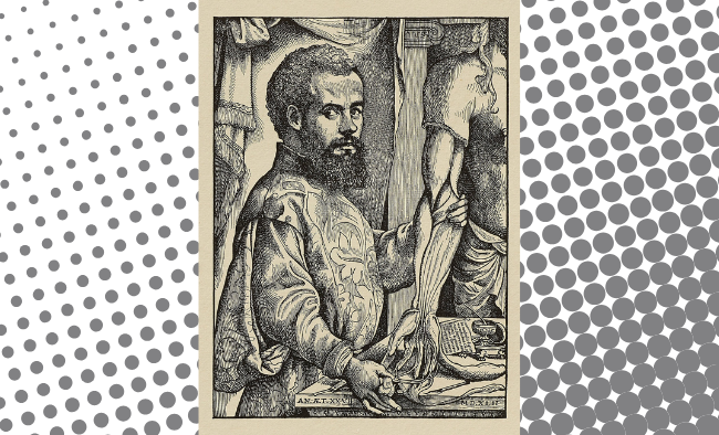
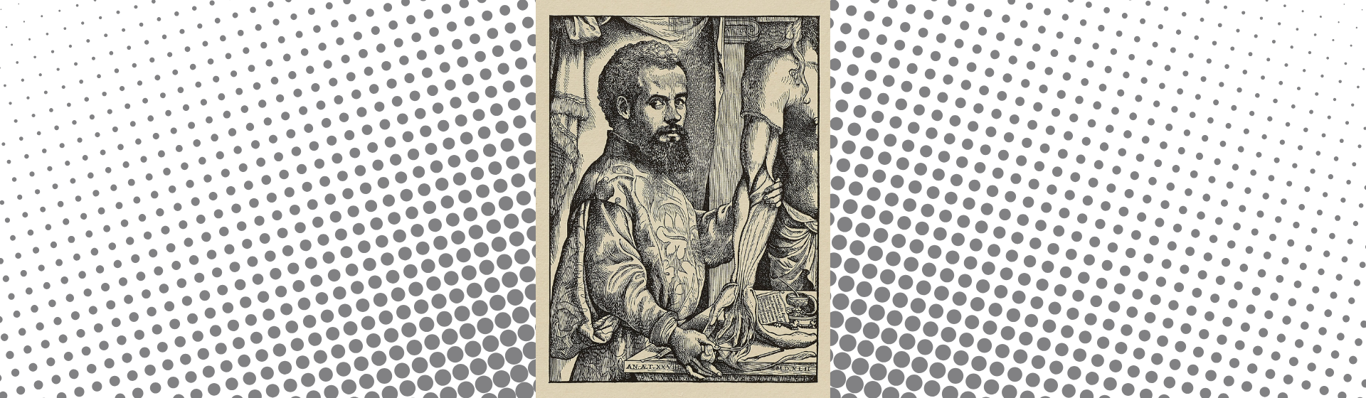
Andreas Vesalius was born in Brussels in 1514 and became one of the central figures in the history of medicine (Downs, 1978). His great work, De humani corporis fabrica libri septem, usually known simply as the Fabrica, was published in Basel in 1543 by Johannes Oporinus. The title can be translated as On the Fabric of the Human Body in Seven Books, and the architectural metaphor in the word fabric is significant. Vesalius presented the body as a structure to be examined, understood, and described through careful observation (Nutton, 2024).
Before Vesalius, European medical education relied heavily on ancient authorities, especially Galen. Galen’s writings were foundational, but many of his anatomical conclusions were based on animal dissection rather than human dissection. Vesalius did not simply repeat what he had inherited. He placed direct observation of the human body at the center of anatomical study, using dissection as both a research method and a teaching practice. That shift helped make anatomy a discipline grounded in evidence rather than solely in textual authority.
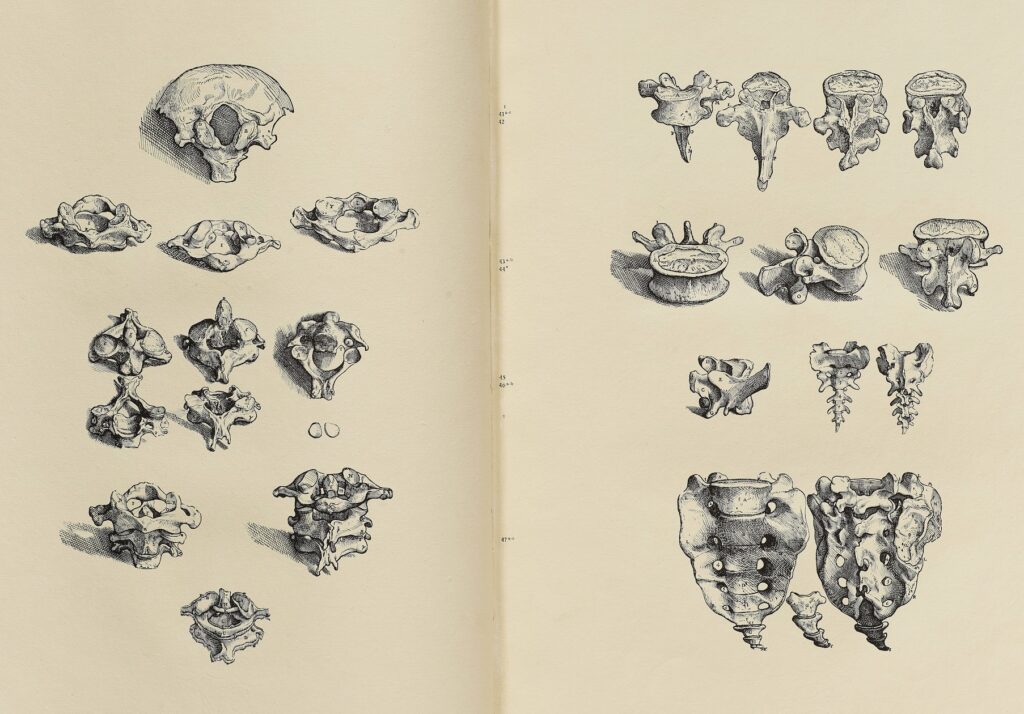
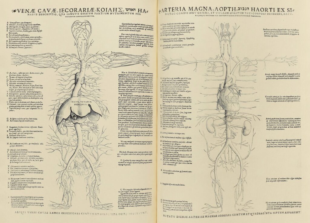
The Fabrica was revolutionary not only because of its scientific argument, but also because of its form as a printed book. It joined detailed anatomical description with more than 200 woodcut illustrations of extraordinary artistic and technical quality. The images were not decorative additions to the text. They were essential to the work’s authority. They allowed readers to see what Vesalius described, and they helped make anatomy into a visual discipline. The precise attribution of the illustrations remains complicated, though the images have long been associated with artists connected to the workshop of Titian, including Jan Stephan van Calcar (Nutton, 2024). Whatever the exact division of labor, the book depended on collaboration among anatomist, artist, woodblock cutter, printer, and publisher.
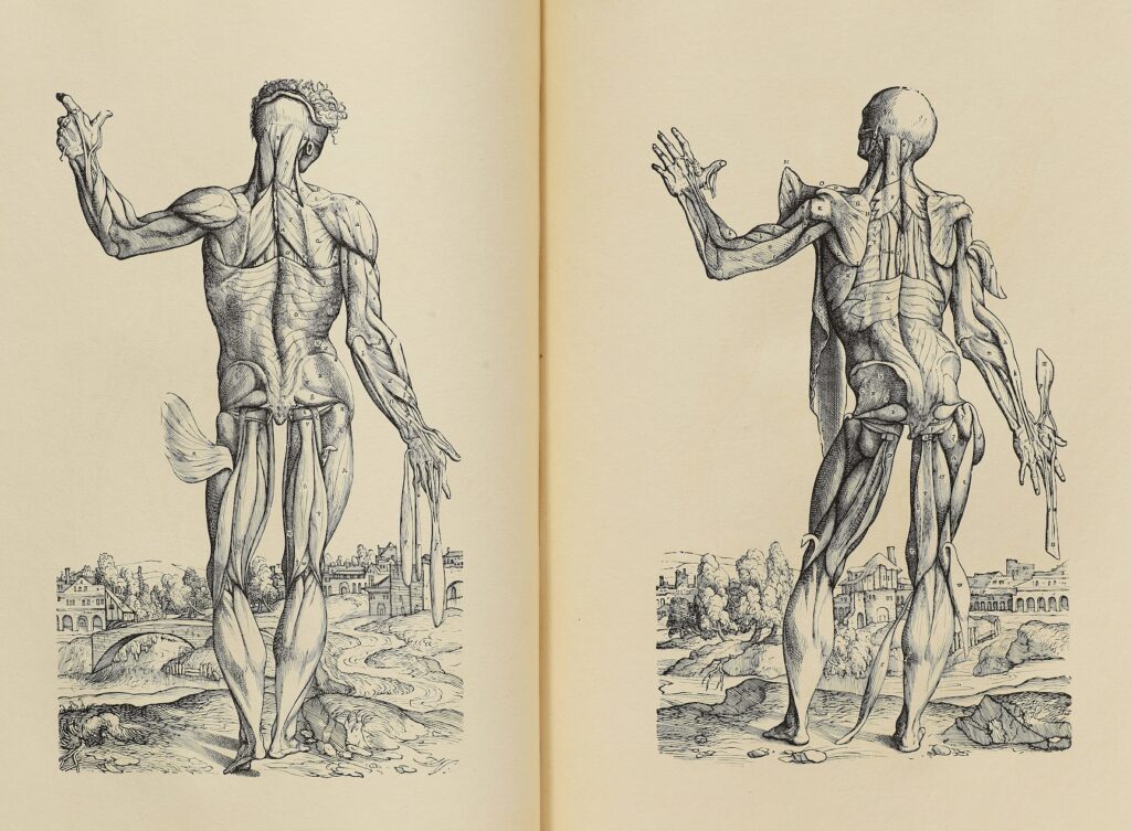
The full-body muscle figures are among the most memorable images in the history of medical illustration. Rather than presenting dissected bodies as static specimens, the figures occupy landscapes and assume expressive poses. Some seem to walk, lean, point, or contemplate their own exposed anatomy. To modern viewers, this can feel unsettling or even surreal, but that tension is part of the power of the images (Kusukawa, 2012). Vesalius’s figures are scientific illustrations, but they are also works of Renaissance visual culture. They insist that anatomy is about the body as an object of study, while never fully separating it from art, mortality, and the human condition.
The 1934 Icones Anatomicae at HPU
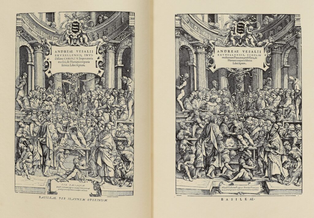
The Vesalius volume now held by HPU Libraries Archives & Special Collections is not a sixteenth-century copy of the Fabrica. It is, however, an important twentieth-century edition directly connected to the survival of Vesalius’s images.
Published in 1934 by the New York Academy of Medicine and the University Library of Munich, Icones Anatomicae was printed by the Bremer Presse in Munich. The edition reproduced Vesalius’s anatomical illustrations, with many images printed from surviving original woodblocks that had been preserved in Munich. HPU’s copy retains its original Frieda Thiersch binding, with vellum spine and corners, gilt titling, deckled handmade paper, and original publisher’s box. It also includes the explanatory Characterum Indices and the surviving plate suite.
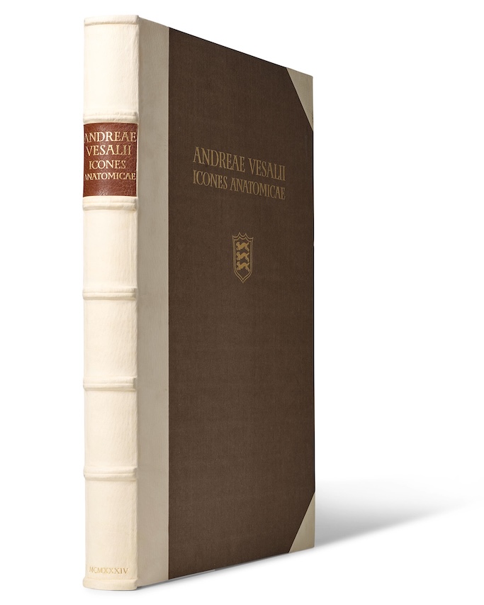
The survival of this edition is especially important because many of the original Vesalian woodblocks were later destroyed during World War II. For that reason, Icones Anatomicae is more than a deluxe facsimile. It preserves impressions from blocks tied to one of the landmark achievements in the history of anatomical illustration. It is also a major achievement in twentieth-century bookmaking, uniting Renaissance image-making with the ideals of the fine press movement.
The physical qualities of the book matter. Handmade paper, vellum, deckled edges, letterpress printing, and original binding all shape how the work is experienced. In a digital age, it can be easy to think of images as detachable from the objects that carry them. This volume reminds us that images also have material histories. They are printed, handled, stored, rebound, boxed, damaged, repaired, donated, cataloged, and displayed. Those histories are part of what Archives & Special Collections preserves.
From woodcut to modern atlas
The modern Thieme Atlas of Anatomy in the display serves many of the same broad purposes as Vesalius’s images, but it speaks a different visual language. Rather than staging the body as a series of dramatic figures, the modern atlas organizes anatomy for clarity, efficiency, and clinical application. Muscles, nerves, vessels, bones, and organs are separated visually. Color helps distinguish systems. Labels, cross-sections, regional views, tables, and clinical notes guide students through complex information.
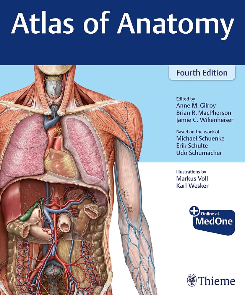
This shift reflects changes in both medicine and design. A sixteenth-century anatomy book had to persuade its readers that direct observation could challenge inherited authority. A twenty-first-century atlas must help students master enormous amounts of information and apply that knowledge in laboratories, simulations, clinics, and patient care settings. Vesalius needed the authority of the dissecting room, the workshop, and the printing press. A modern atlas needs the visual discipline of graphic design, standardized terminology, and pedagogical sequencing.
Yet the comparison also shows continuity. Anatomical illustration is never simply a matter of copying the body in that every image makes choices. What should be included? What should be simplified? Which structures should be emphasized? Should the viewer see the body as a whole human figure, a specimen, a region, a system, a layer, or a clinical problem? How should color, scale, perspective, and labels shape the reader’s attention? These are scientific questions, but they are also artistic and design questions.
The National Library of Medicine’s Dream Anatomy exhibition makes this point especially well, tracing a long history of anatomical images that are informative, beautiful, strange, unsettling, and imaginative. Across centuries, the body has been represented through woodcut, engraving, hand coloring, lithography, photography, X-ray imaging, and digital imaging. The movement from Vesalius to a modern atlas is therefore not a simple story of old images becoming more accurate. It is a story of changing technologies, changing audiences, and changing ideas about what medical images are supposed to do.
Why this matters at HPU
This display is especially relevant to High Point University because it sits at the intersection of several areas of teaching and learning across campus.
For students in the Congdon School of Health Sciences, anatomy is foundational. HPU’s health sciences programs include Exercise Science, Health and Wellness, Physical Therapy, Physician Assistant Studies, Athletic Training, Biomedical Sciences, Doctor of Medical Science, and Healthcare Administration. The school emphasizes interprofessional education, evidence-based clinical practice, community engagement, and preparation for patient-centered health care. Its facilities include the Center for Medical Simulation, the Human Gross Anatomy Lab, the Human Biomechanics and Physiology Lab, the Pro Bono Physical Therapy Clinic, Targeted Enhanced Athletic Movement, and the Virtual Reality and Clinical Gait Analysis Laboratory.
A display like this invites health sciences students to think historically about the tools they use every day. Anatomy is not only a set of facts to memorize. It is a tradition of observation, representation, teaching, revision, and ethical responsibility. Vesalius’s work reminds us that medical knowledge advances when people look closely, test assumptions, and communicate what they see in forms others can understand.
The display also speaks directly to students and faculty in the David R. Hayworth School of Arts and Design. The school houses programs in Studio Art, Graphic Design, Fashion Merchandising, Interior Design, Music, Theater, and Dance, along with minors including Art History, Museum Studies, Photography, and Visual Merchandising Design. Anatomical illustration offers a powerful example of how visual communication works. An anatomical image must be accurate enough to teach, clear enough to guide the eye, and compelling enough to hold attention. It must balance beauty and utility, complexity and comprehension.
For graphic design students, a modern anatomy atlas is a lesson in information design. It uses hierarchy, color, labels, spacing, sequence, and repetition to make complicated material usable. For studio art students, Vesalius shows the enduring relationship between drawing, observation, proportion, and the human figure. For museum studies and visual merchandising students, the display raises interpretive questions about how to present rare books safely, how to write for public audiences, and how to use a small exhibit to create curiosity across disciplines.
Other programs have a stake in this conversation as well. Students in the Wanek School of Natural Sciences encounter anatomy through biology, physiology, neuroscience, evolution, and laboratory observation. Students in the Teresa Caine School of Nursing and the Fred Wilson School of Pharmacy rely on anatomical and physiological knowledge as part of patient-centered care. Students in the Workman School of Dental Medicine and the School of Optometry study specialized structures of the head, mouth, eye, and nervous system. Students in Communication can ask how scientific images persuade, while students in History and Humanities can examine how books, bodies, and knowledge have been shaped by culture, technology, and institutions.
Rare books as teaching objects
One goal of Archives & Special Collections at HPU Libraries is to make rare and distinctive materials more visible, more useful, and more connected to teaching. A book like Icones Anatomicae can support instruction in the history of medicine, biology, graphic design, art history, book history, museum studies, and humanities seminars on visual evidence. It can also help students understand that knowledge is not abstract. It is produced through tools, materials, institutions, and people.
The modern atlas is equally valuable in this display because it keeps the conversation from becoming purely historical. Today’s textbooks, eBooks, databases, simulations, and digital learning tools belong to the same long story. Students who use contemporary anatomical resources are participating in a tradition that extends back centuries. The technologies have changed, but the central challenge remains familiar: how can the complexity of the human body be transformed into images that can be studied, remembered, taught, and applied?
By bringing these books together, Archives & Special Collections can support object-based learning across campus. Students can encounter a rare book not as a sealed treasure, but as an active teaching object. They can ask how it was made, why it mattered, how it survived, and what it can still teach us. Faculty can use it to build conversations across disciplines, connecting human anatomy to image-making, pedagogy, ethics, preservation, and the history of print.
Seeing the body, seeing the book
The pairing of Vesalius’s Icones Anatomicae with a modern Thieme Atlas of Anatomy is a small display with a large story. It shows how anatomical illustration has moved from hand-cut woodblocks to full-color teaching atlases, from Renaissance dissection theaters to modern simulation labs, and from dramatic whole-body figures to clinical diagrams and layered visual systems.
It also shows continuity. Across centuries, anatomy has required careful observation, skilled image-making, teachers, students, artists, printers, publishers, donors, collectors, and libraries. Most of all, it has required the belief that seeing clearly can deepen understanding.
That is what makes these books so compelling together. One preserves the visual world of Renaissance anatomy through a twentieth-century fine press edition. The other represents the modern visual language of health sciences education. Both ask us to look closely at the human body, and at the books that have helped generations understand it.
References
- Downs, R. B. (1978). Books that changed the world (2d ed). American Library Association.
- Gilroy, A. M., MacPherson, B. R., & Ross, L. M. (2010). Atlas of anatomy. Thieme.
- Kusukawa, S. (2012). Picturing the book of nature: Image, text, and argument in sixteenth-century human anatomy and medical botany. University of Chicago Press.
- National Library of Medicine. (n.d.). Dream Anatomy. National Institutes of Health. https://www.nlm.nih.gov/exhibition/dream-anatomy/index.html
- National Library of Medicine. (n.d.). Historical Anatomies on the Web: Andreas Vesalius. National Institutes of Health. https://www.nlm.nih.gov/exhibition/historicalanatomies/vesalius_home.html
- Nutton, V. (2024). Andreas Vesalius and his Fabrica, 1537-1564: Changing the world of anatomy. Palgrave Macmillan.
- Vesalius, A. (1934). Icones anatomicae. New York Academy of Medicine and University Library of Munich. Printed by the Bremer Presse.
- Vesalius, A. (1950). The illustrations from the works of Andreas Vesalius of Brussels (J. B. d. M. Saunders & C. D. O’Malley, Eds.). Dover Publications.